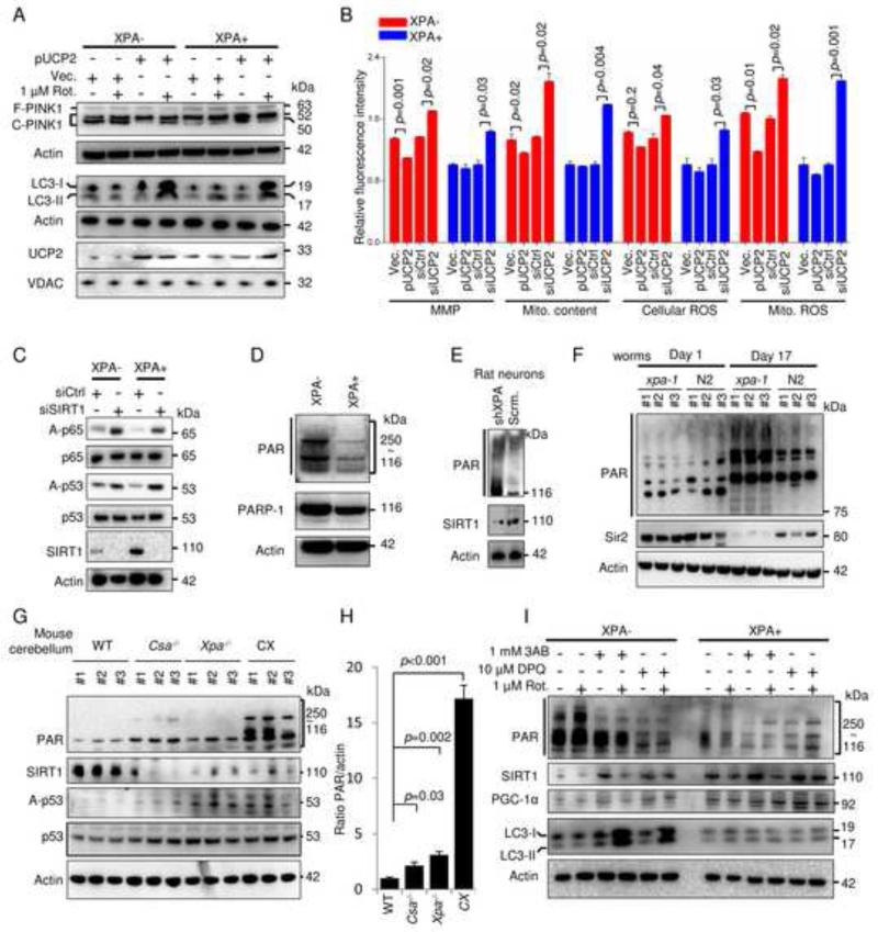
Figure 5. The NAD+-SIRT1-PGC1-α axis regulates the expression of UCP2 which can rescue the mitophagy defect in XPA-cells.
(A) XPA− and XPA+ cells were transfected with pUCP2 or an empty vector for two days, and then treated with 1 μM rotenone or vehicle for 24 h, followed by immunoblot for proteins as indicated. (B) XPA− and XPA+ cells were transfected with pUCP2, vector, control siRNA or siRNA targeting UCP2 for two days, subsequently indicated parameters were analysed by flow cytometry (means ± S.E.M., n=3). (C) Immunoblot of XPA− and XPA+ cells after transfection with control siRNA or siRNA targeting SIRT1 (siSIRT1). (D) Representative immunoblot of PAR and PARP-1 in XPA− and XPA+ cells. (E) Immunoblot showing expression of PAR and SIRT1 in primary rat neurons with shRNA XPA knockdown or control shRNA. (F) Immunoblot of whole cell extracts from young (day 1) and old (day 17) xpa-1 mutant and WT (N2) nematodes. (G) Immunoblot of cerebellar protein levels in 2-week old WT, Csa−/−, Xpa−/− or Csa−/−/Xpa−/− (CX) mice and quantification in (H) (means ± S.D., n=3). (I) Immunoblot of XPA− and XPA+ cells treated with two PARP inhibitors, 3AB (1 mM) and DPQ (10 μM), for 12 h, followed an additional 24 h treatment in the presence of 1 μM rotenone or vehicle. See also Figure S5.

