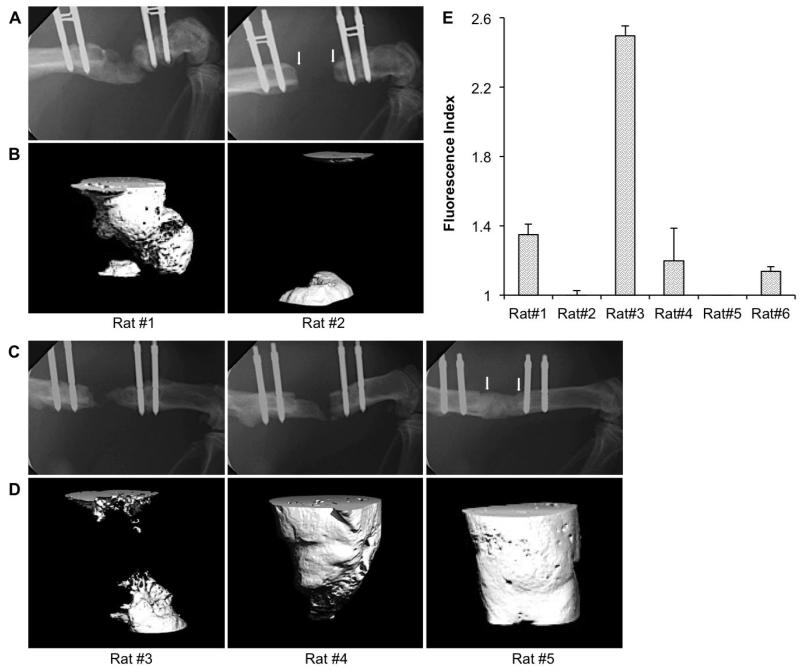
Fig. 1.
Bone healing in athymic rats. Radiologic findings: Representative images of X-ray (A, C) and corresponding high resolution μCT (B, D) taken at 8 weeks, showing the typical range of bone healing efficacy in athymic rats receiving grafts of genetically modified sheep fat (A, B) or muscle (C, D). Arrows in the X-ray images of rats # 2 and 5 show the edges of the defect. Immunologic findings: Rat anti-sheep IgG xenoantibody responses in six different rats at 8 weeks following sheep muscle implantation (E). Samples were run in triplicate.

