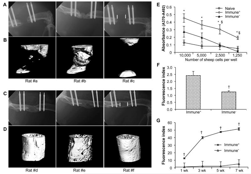
Fig. 2.
Bone healing in immunosuppressed Fischer 344 rats. Radiologic findings: Representative X-rays (A, C) and corresponding high resolution μCT (B, D) scans taken at 8 weeks, showing new bone formation in rats receiving grafts of genetically modified sheep fat (A, B) or muscle (C, D). Immunologic findings: (E) Proliferation of splenocytes recovered from rats receiving grafts of genetically modified sheep muscle. Data are presented as mean values × 103 ± SEM; * and $ denote a significant difference (p < 0.05) relative to naïve control group and immunocompetent group (Immune+) group, respectively. (F) Rat anti-sheep IgM responses at 8 d. (G) IgG xenoantibody responses over time following sheep muscle implantation. † indicates a significant difference when compared to control (Immune+) group at respective time-points (p < 0.05). For the immunosuppressed (Immune−) group, no significant difference existed between different time-points. All immunoassays were run in triplicate.

