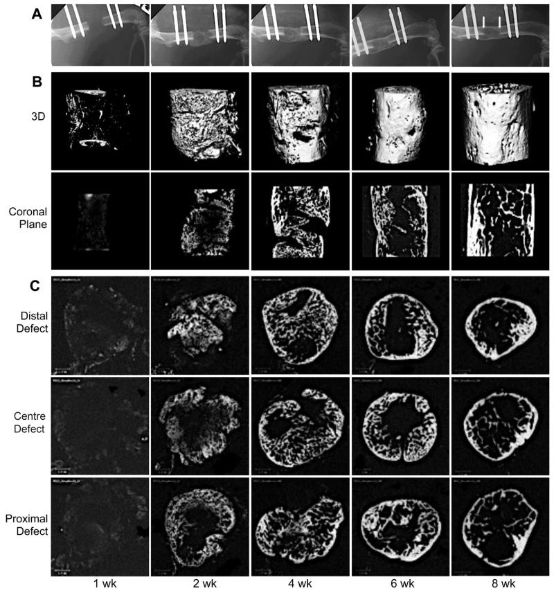
Fig. 3.
Serial radiographs and μCT scans of femoral defects treated with genetically modified sheep muscle. Criticalsized segmental femoral defects in Fischer F344 rats were implanted with Ad-BMP2 transduced sheep muscle grafts and transiently immunosuppressed. X-rays (A) were taken weekly and the defects were then harvested for μCT scans (B, 3-dimensional and coronal images; C, cross sectional images) at progressive time-points after surgery. Arrowheads in panel A, 8 weeks indicate the original defect boundaries. Scale bar = 1 mm.

