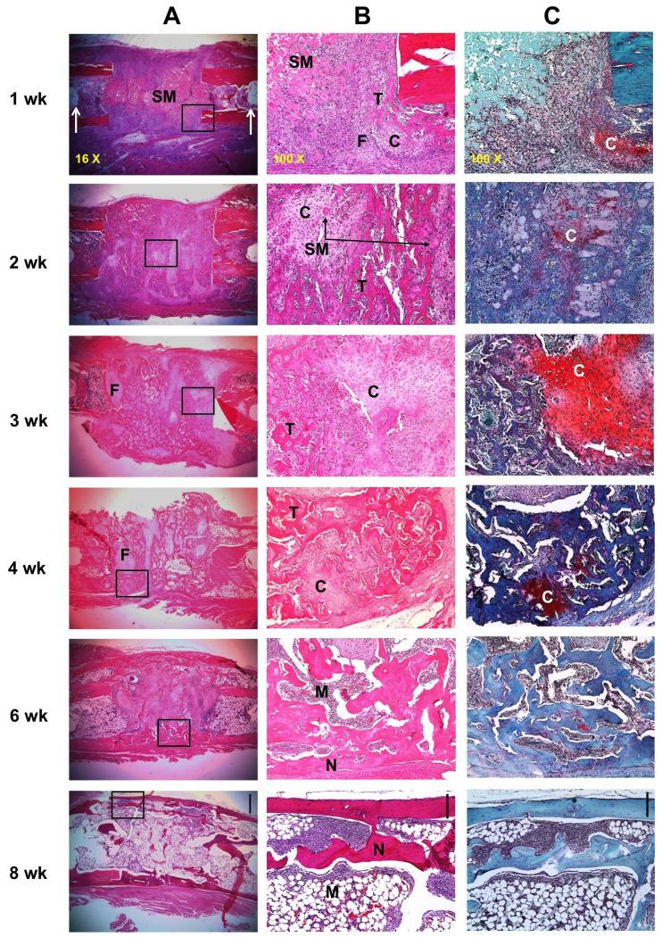
Fig. 5.
Histological appearance of defects during healing in transiently immunosuppressed rats. Ad.BMP-2 transduced sheep muscle grafts were implanted into defects in Fischer 344 rats and harvested for histology at different time-points. Rats were immunosuppressed for 3 weeks. (A) Representative images of sections stained with haematoxylin and eosin under low magnification (scale bar in 8 week panel = 1 mm). The boxed regions shown in panel A are shown at high magnification (scale bar = 0.2 mm) (B) and stained with safranin orange-fast green (scale bar = 0.2 mm) (C). Arrows in panel A, 1 week, indicate the pin holes of the external fixator. N indicates new cortical bone, C indicates cartilage, T indicates trabecular bone, M indicates bone marrow, SM indicates sheep muscle tissue, and F indicates fibrous tissue.

