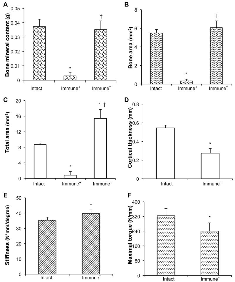
Fig. 6.
Physical properties of the defects 8 weeks after implantation of genetically modified, sheep muscle grafts. (A) Bone mineral content, (B) Bone area, (C) Total callus area, (D) Cortical thickness, (E) Stiffness, (F) Strength measured as maximum torque. * denotes significant difference (p < 0.05) when compared to intact contralateral femur group; † indicates significant difference (p < 0.05) relative to immunocompetent (Immune+) group. It was not possible to perform mechanical testing on non-immunosuppressed (Immune−) samples and these had no cortices.

