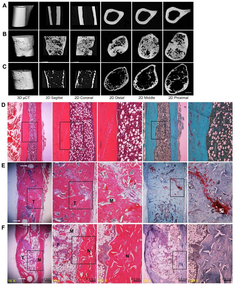
Fig. 7.
Comparison of new bone formed in athymic rats and immunosuppressed Fischer 344 rats. Representative μCT images of normal diaphyseal femur (A), and repair bone formed 8 weeks after implantation of genetically modified sheep muscle in athymic rats (B) or in transiently immunosuppressed Fischer 344 rats (C). Representative histological images, shown at progressively higher magnification from left to right, display normal diaphyseal femur (D) and repair bone formed in athymic (E) and immunosupressed (F) rats. Sections were stained with H&E and safranin orange-fast green. Arrows in panels E and F, left hand panels, indicate the pin holes of external fixator. N indicates new cortical bone within the defects, M indicates bone marrow, T indicates trabecular bone, and F indicates fibrous tissue.

