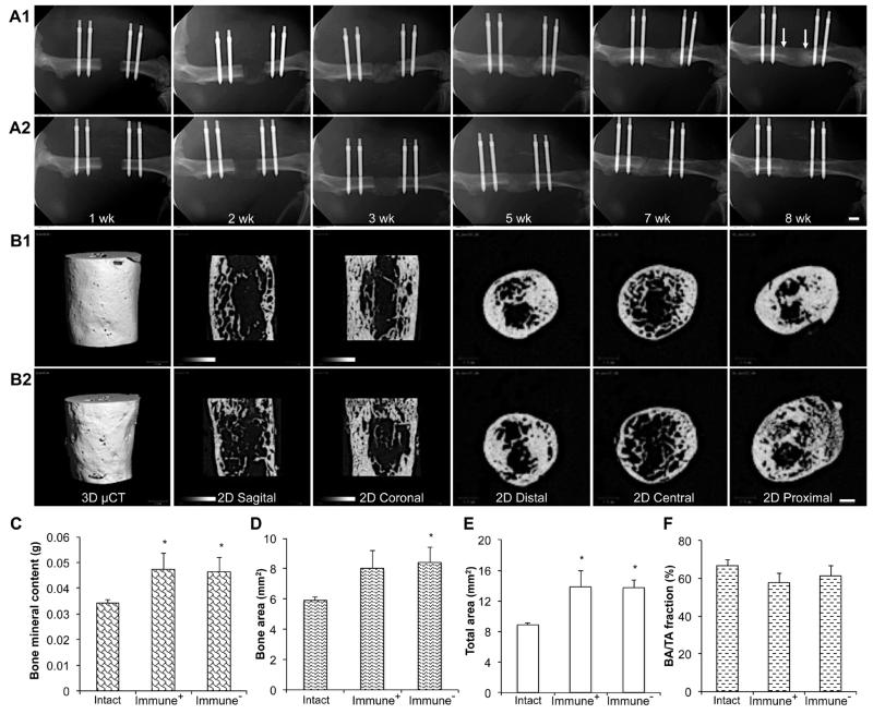
Fig. 8.
Effects of transient immunosuppression on bone healing in response to rhBMP-2. Radiology: Serial X-ray images (A1, A2) and μCT images at 8 weeks (B1, B2) of defects during healing in the absence (A1, B1) or presence (A2, B2) of immunosuppression. Arrows in panel A1, 8 weeks, indicate the original defect boundaries. Scale bars = 1 mm. Physical properties: Bone mineral content (C), Bone area (D), Total callus area (E), and Bone area/Total area or Callus size (F, BA/TA, %).

