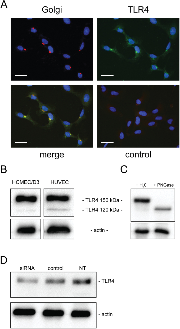
Fig. 4.
TLR4 expression in endothelial cells. (A) TLR4 co-localizes with the Golgi apparatus. Fluorescent staining of Golgi apparatus (red), TLR4 (green) and nuclei (blue) on HCMEC/D3 cells (not confluent) after fixation with PFA 2% and permeabilization with saponin 0.2%. Negative control is obtained after staining with the secondary antibodies only. Pearson’s correlation coefficient Rr = 0.67 and Mander’s overlap coefficient R = 0.75. Scale bar: 20 µm. (B) TLR4 is expressed on HCMEC/D3 and on HUVECs. Cells were lysed with the Tris–HCl 50mM, pH 7.5, NaCl 150mM buffer containing 1% NP40. The presence of TLR4 in the whole extracts is then analyzed by western blotting. (C) TLR4 heavy form (150kDa) leads to a light form (120kDa) after treatment with PNGase. Whole extracts, prepared with a lysis buffer containing only NP40, were treated with PNGase, or water as a negative control, for 2h and then analyzed by western blotting. (D) TLR4 expression on HCMEC/D3 cells after double transfection with siRNAs targeting TLR4 or control siRNAs; cells were lysed with the Tris–HCl 50mM, pH 7.5, NaCl 150mM buffer containing 1% NP40. The presence of TLR4 in the whole extracts was then analyzed by western blotting and quantified with ImageJ software. NT: not transfected.

