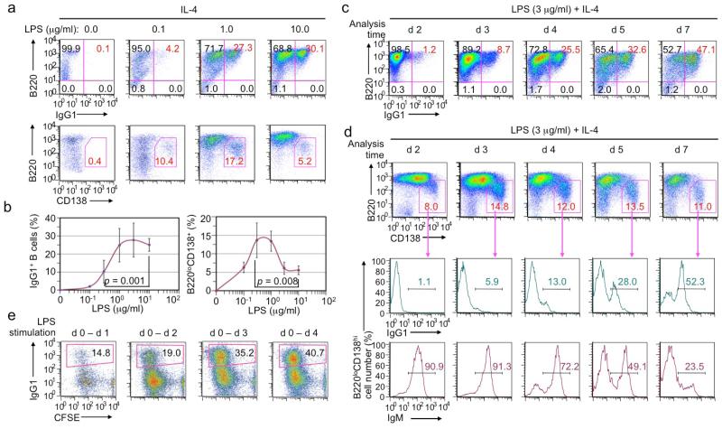
Figure 2.
LPS induces B cells to undergo CSR and plasma cell differentiation in vitro in a dose- and time-dependent manner (flow cytometry analysis). (a, b) CSR to IgG1 in B cells stimulated with LPS at the indicated concentrations plus IL-4 for 4 d (a, top panels), and formation of B220loCD138hi plasma cells in the same cell culture (a, bottom panels). Mean and s.d. of data from four independent experiments were depicted in (b). p values calculated by paired student t test. (c) CSR to IgG1 in B cells stimulated with LPS plus IL-4, starting at d 0, and harvested at d 2, d 3, d 4, d 5 and d 7, as indicated. (d) Formation of B220loCD138hi plasma cells (top panels) as well as the proportion of IgG1+ (middle panels) and IgM+ (bottom panels) within those plasma cells in cultures of B cells stimulated with LPS plus IL-4, starting at d 0, and harvested at d 2, d 3, d 4, d 5 and d 7, as indicated. (e) CSR to IgG1 in B cells stimulated with LPS plus IL-4 for the period indicated, pelleted to remove LPS, and then cultured in RPMI-FBS only until being harvested at d 4. Data in (a, c, d, e) are representative of three independent experiments.

