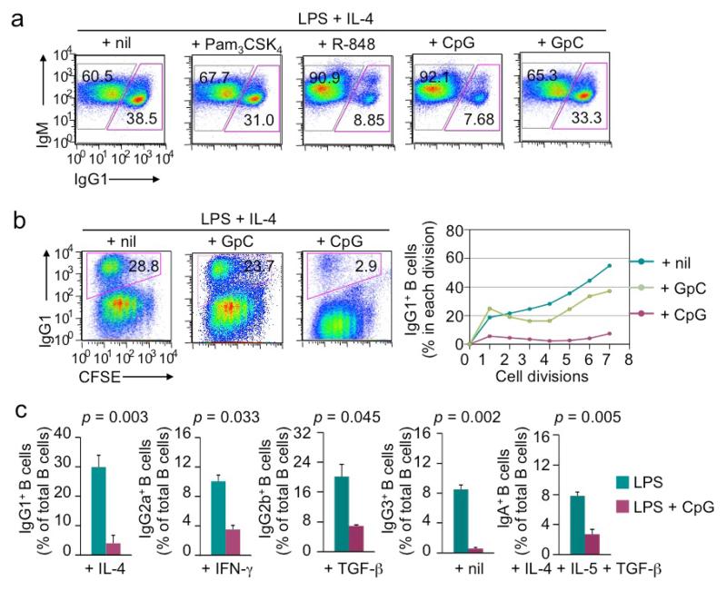
Figure 5.
LPS-induced CSR is inhibited by CpG and R-848 in the absence of BCR crosslinking (flow cytometry analysis). (a) The proportion of switched IgG1+IgM− B cells and IgM+IgG1− unswitched B cells in B cells after stimulation with LPS (3 μg/ml) plus IL-4 in the presence of nil, Pam3CSK4, R-848, CpG or GpC for 4 d. (b) CSR to IgG1 in CFSE-labeled B cells stimulated with LPS (3 μg/ml) plus IL-4 in the absence or presence of GpC (0.3 μM) or CpG (0.3 μM; left panels) for 4 d and depiction of the proportion of switched IgG1+ cells in B cells that had completed each cell division (right panels). (c) CSR to different IgG isotypes or IgA in B cells stimulated with LPS (3 μg/ml) plus appropriate cytokines (as indicated) in the absence or presence of CpG (0.3 μM) (mean and s.d. of data from three independent experiments; p values calculated by paired student t test). Data in (a) and (b) are representative of three independent experiments.

