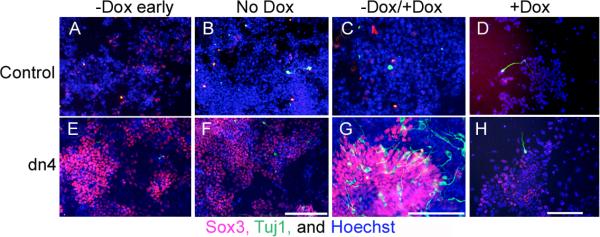
Figure 3. Regulated expression of the dnTcf4 protein promotes neural differentiation of ESC in a monolayer assay.

Immunohistochemical localization of cell type restricted antigens in Control (A-D) and dnTcf4 (E-H) ESC grown in monolayer. When the transgene was induced and Wnt signaling was abrogated for 4 (-Dox early; E) or 6 days (No Dox; F) of differentiation there was widespread expression of the neural precursor marker Sox3 (red, Cy3 secondary) compared with controls (A,B), but was little expression of the neuronal marker βIII tubulin (Tuj1 antibody, green, FITC secondary; A,B, E,F). Reactivation of Wnt signaling during the last 3 days of differentiation (-Dox/+Dox; G) resulted in a significant increase in the conversion of Sox3 positive precursors to immature neurons, compared with Controls (C). There was little neuronal differentiation when Control or dn4 cells were grown continuously in doxycycline for 6 days (+Dox; D,H). Nuclei were stained with Hoechst (blue). Scale bar = 200 μM.
