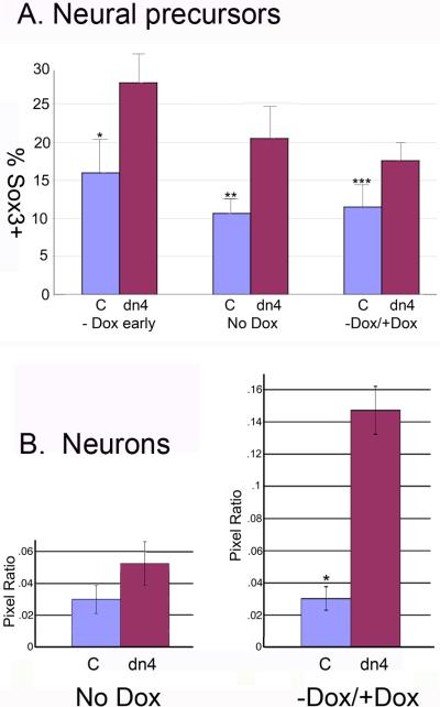
Figure 4. Regulated expression of the dnTcf4 protein significantly increases neural and neuronal differentiation of ESC in a monolayer assay.
A. FACS analysis of neural precursors (Sox3 +cells) following induction of the dnTcf4 transgene. Cells were cultured for 3 days without Dox (-Dox early), 6 days without Dox (No Dox), or 3 days without Dox followed by 3 days with Dox (-Dox / +Dox). The number of Sox3 positive cells was significantly increased in dnTcf4 (dn4) expressing cells compared to control cells (C) at all time points. *p≤0.001, p**≤0.01, p***≤0.05, Student's t test. The graphs represent averages of 5 experiments with 3 replicates/experiment.
B. The number of neurons in control and dnTcf4 ESC grown in monolayer culture was quantified using NIH Image J. IHC localization of βIII tubulin was carried out in cultures in which Wnt signaling was abrogated during the entire culture period (No Dox) or when signaling was abrogated only during the first half of the culture period (-Dox / +Dox). The average number of βIII tubulin pixels was divided by the average number of Hoechst pixels in 10 fields per well, 3 replicates/experiment, in 4 biological experiments (n=120 fields/group). There was a significant increase in neuronal differentiation only when Sox3 positive precursors were allowed to complete differentiation in the presence of Wnt signaling during the last half of the culture period (-Dox / +Dox). *p≤0.003 Student's t test.

