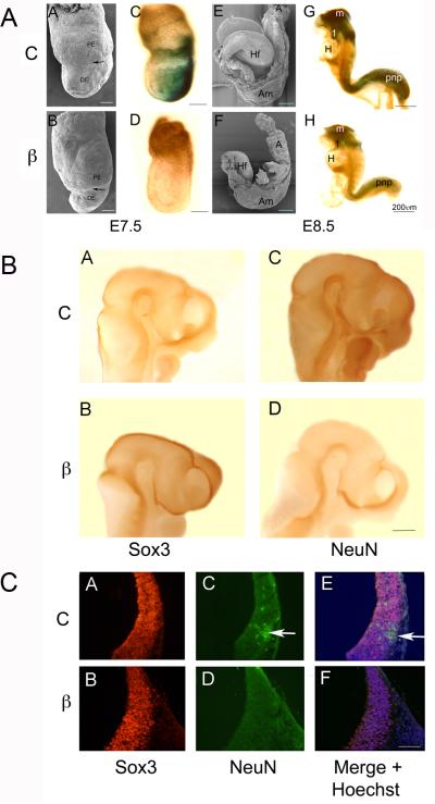
Figure 7. Wnt signaling is required in the embryo for the conversion of Sox3 neural precursors to NeuN positive neurons.
A. Compared with control (C) embryos (A,C,E,G), embryos (β) exposed to shRNA targeting β-catenin (B,D,F,H) exhibit defects in gastrulation, axis elongation, and neural differentiation. Side views of E7.5 and 8.0-8.5 embryos examined using SEM (AB, EF) or stained with Xgal to identify sites of β-galactosidase expression and Wnt signaling (CD, GH). The arrows in A and B indicate the boundary between primitive endoderm (PE) and definitive endoderm (DE) that largely replaces the PE at gastrulation. The proximal-distal displacement of the boundary is an indication of the progress of gastrulation movements. The control embryo (A) has expanded in both the proximal-distal (P-D) and anterior-posterior (A-P) axes compared to the β-catenin shRNA exposed embryo (B). In control embryos (C) Wnt signaling is strong in the posterior primitive streak (PS) compared with shRNA exposed embryos (D) where signaling is nearly abrogated. By day 8 in control embryos (E) the neural folds are elevating and later embryos (G) are beginning the process of adopting the fetal C shape. β-galactosidase is expressed at high levels in the midbrain (m), first branchial arch (1), and posterior neuropore (pnp) in control embryos (G), but is strikingly down-regulated in β-catenin shRNA treated embryos (H). Anterior is to the left in each figure, Am=amnion, A=Allantois, Hf=headfold, H=heart. SBs = 200μm
B. Side views of control (A, C) and embryos exposed to shRNA against β-catenin (B, D). Anterior is to the right in A-D. Whole mount IHC was carried out to compare Sox3 positive neural precursors (A, B) and NeuN positive neurons (C, D). By E10.5 in control embryos there were more NeuN positive neurons (C) and fewer Sox3 positive precursors (A) compared with β-catenin shRNA exposed embryos (B, D). Embryos exposed to β-catenin shRNA also exhibited increased midbrain mesenchyme and abnormal positioning of midbrain flexure.
C. When similar embryos were sectioned, there was widespread expression of Sox3 in both control and β-catenin shRNA exposed embryos (A, B) while NeuN was observed in differentiating neurons in control embryos (C, arrow) but not in embryos exposed to β-catenin shRNA (D). E and F are overlays of AC+BD with Hoechst staining of nuclei. SB=200μ

