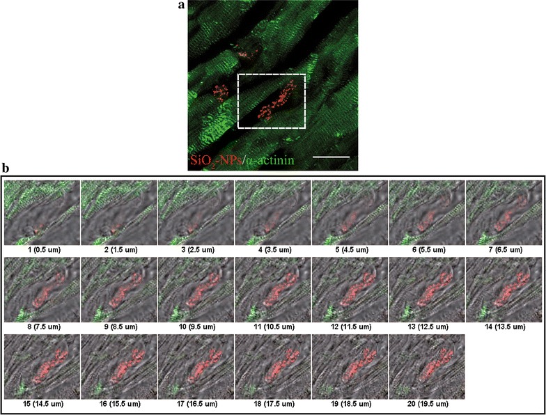
Fig. 7.

Ultrastructural analysis of labelled hMSCs engrafted inside ventricular tissue. Representative confocal reconstruction of a normal ventricle. a Superposition of sarcomeric α-actinin staining (green) and SiO2-NPs internalized in hMSCs (red). Magnification 100×, scale bar 20 µm. b Consequent slices along the Z-axis from the subset in A (dashed perimeter), to display morphological rearrangement of a hMSC between cardiac fibers; superposition of α-actinin (green), SiO2-NPs (red) and correspondent bright field to reveal cell body
