
Figure 2. Expression of HDAC 1 and 2.
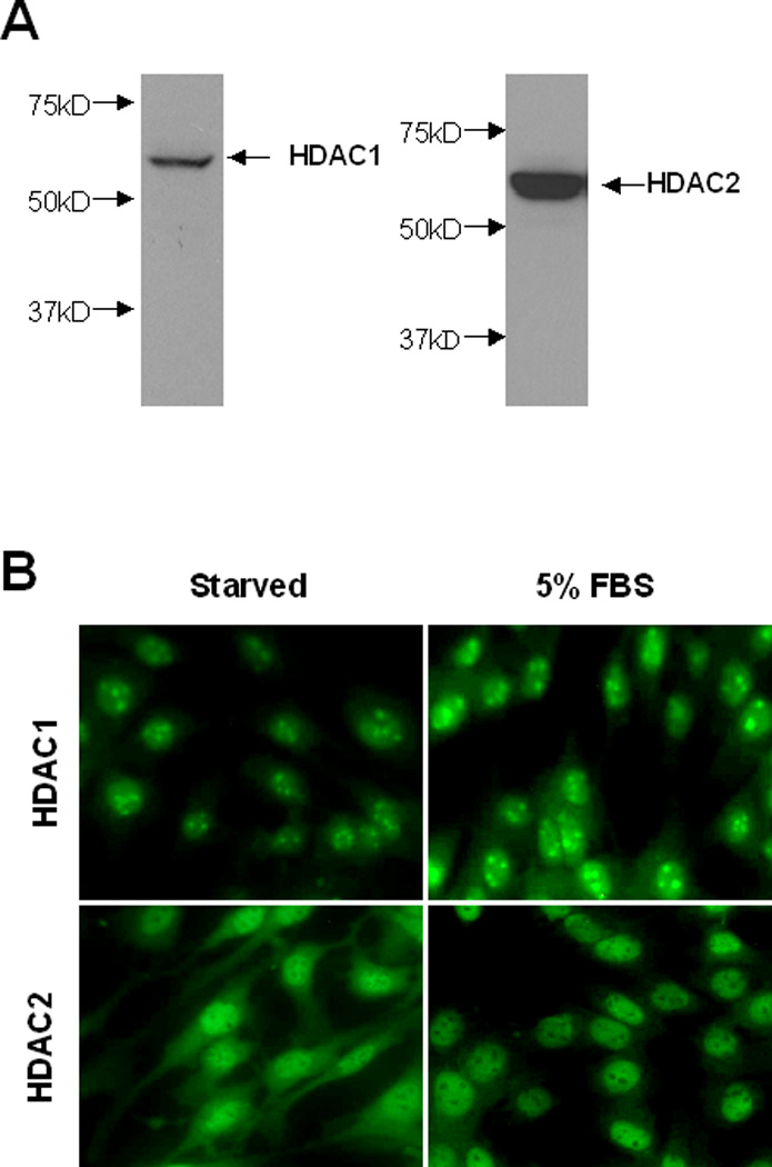
(A) Cultured NRK-49F cells were harvested and cell lysates were subjected to immunoblot analysis with antibodies against HDAC1 and HDAC2. Representative immunoblots from 3 or more experiments are shown (1X200). (B) Cultured NRK-49F cells were seeded on the cover slips and then starved for 24 h with 0.5% FBS. After treatment with 5% FBS for an additional 1 h, cells were harvested for staining with antibodies to HDAC1 or HDAC2.