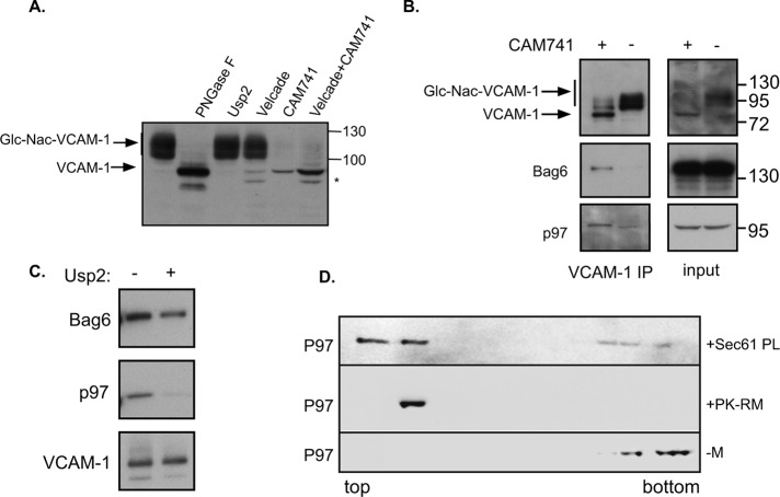
FIGURE 1:
(A) VCAM1-HA was transfected into cells, and 48 h posttransfection, VCAM1 content was evaluated by HA immunoblots. Cell lysates were analyzed directly or after in vitro PNGase or Usp2 treatment. Where indicated, cells were treated overnight with Velcade (100 nM) and/or CAM741 (250 nM) as indicated. Asterisk marks an additional LMW band of VCAM-1. Glycosylated VCAM-1 is labeled Glc-Nac-VCAM-1. (B) Cells expressing VCAM1 were treated with Velcade in the presence or absence of CAM741 as indicated. Cell lysates were evaluated directly (input) or subjected to a HA IP to evaluate Bag6 and p97 interaction. (C) VCAM-1 IP in the presence of CAM741 and Velcade as in B was subjected to an in vitro deubiquitination by Usp2 as indicated. (D) Recombinant P97 was layered under a discontinuous sucrose gradient in the absence of membranes (–M) or presence of ribosome-stripped PK-RMs or purified reconstituted trimeric Sec61-PLs. After centrifugation, fractions were collected from top to bottom and immunoblotted against p97. The membrane binding of p97 is evident by its flotation together with the membranes to the top of the gradient.

