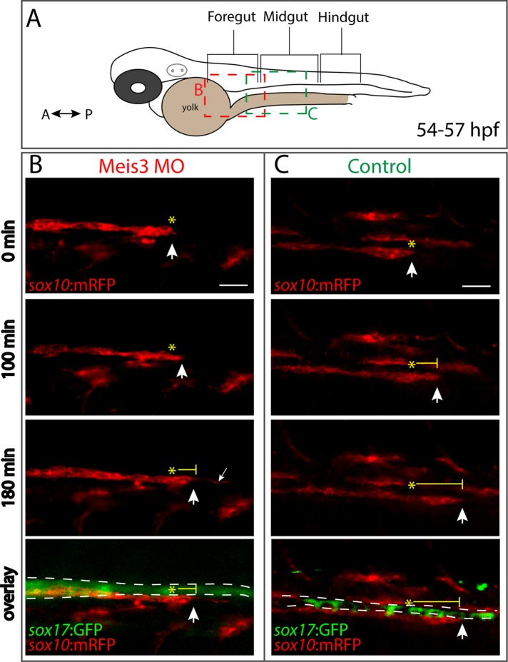
FIGURE 4:
Meis3 is required for efficient migration of enteric neural crest cells along the gut. (A) Schematic depicting where along the gut axis live time-lapse movies were acquired. Colored boxes depict where neural crest streams were observed in Meis3 morphant (red box) and Control embryos (green box). Lateral cropped view of time-lapse stills depicting sox10:mRFP+; sox17:GFP+ (B) Meis3 morphant and (C) control embryos over time during the second day in development (∼54–57 hpf). (B) In Meis3 morphant embryos, sox10:mRFP+ enteric neural crest chains were observed migrating at a rate of 10 μm/h along the posterior end of the foregut, where they migrated 30 μm within 3 h (arrow over time). In Meis3 morphants, the leading edge of the migratory front exhibited membrane protrusions caudally along the gut (small arrow). (C) In control embryos, sox10:mRFP+ enteric neural crest migratory chains were observed migrating at a rate of 23 μm/h along the midgut, where they migrated 70 μm within 3 h (arrow over time). Asterisks mark beginning of migratory front; bottom, merging shows location of sox10:mRFP+ enteric neural crest cells (red) along the sox17:GFP+ gut endoderm (green). Scale bar, 40 μm.

