FIGURE 5:
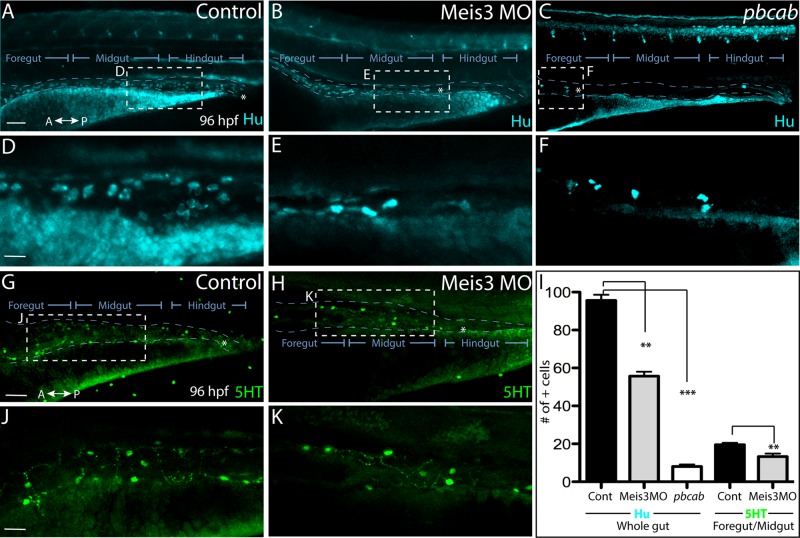
Loss of Meis3 leads to colonic aganglionosis during ENS development. Whole-mount immunohistochemistry against Hu at 96 hpf reveals enteric neurons present throughout the entire length of the gut of (A) control embryos, while they are absent from the hindgut region of (B) Meis3 morphants and present only within the foregut of (C) pbcab-injected embryos. (D) Zoomed-in view from A shows enteric neuron density and morphology in control embryos, whereas zoomed-in views from (E) Meis3 morphants in B and (F) pbcab-injected embryos in C reveal fewer neurons present after loss of Meis3. (G, J) Whole-mount immunohistochemistry against 5HT neurotransmitter in control embryos reveals the presence of terminally differentiated neurons throughout the gut. (H, K) In Meis3 morphants, the presence of 5HT+ terminally differentiated neurons was evident within the foregut and midgut but not the hindgut region, marked by asterisk. (I) Graph quantifying total Hu neurons in control-, Meis3 MO–, and pbcab-injected guts at 96 hpf, as well as total 5HT neurons within the foregut-midgut of control and Meis3 morphant guts. **p < 0.05; ***p < 0.01. Error bars indicate ± SEM. Scale bar, 80 μm (A– C, G, H), 30 μm (D–F, J, K).

