FIGURE 6:
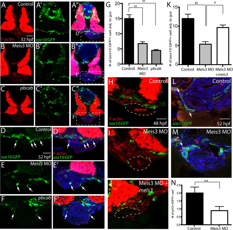
Meis3 function is required for the proper number of enteric neural crest cells around the gut and for their proliferation. By use of immunohistochemistry, enteric neural crest cell numbers were analyzed in (A–A′′) control-, (B–B′′) Meis3 MO–, and (C–C′′) pbcab-injected sox10:GFP+ embryos at 52 hpf. Zoomed-in views of control- (D, D′), Meis3 MO– (E, E′), and pbcab-injected embryos (F, F′) reveal fewer enteric neural crest cells near the gut after loss of Meis3 or Meis function; sox10:GFP (green), F-actin (red), and DAPI (blue). (G) Bar graph showing quantification of enteric neural crest near the gut in control-, Meis3 MO–, and pbcab-injected embryos at 52 hpf. (H–K) Rescue experiment immunohistochemistry and quantification showing that coinjection of meis3 mRNA, which cannot be bound by Meis3 MO, along with Meis3 MO (J) was sufficient to partially rescue enteric neural crest cell number near the gut at 48 hpf (K) when compared with injection of Meis3 MO alone (I, K); sox10:GFP (green), F-actin (red). (L) Control immunohistochemistry against the mitotic marker pH3 at 52 hpf and in (M) Meis3 MO–injected embryos; sox10:GFP (green), pH3 (red), and DAPI (blue). (N) Bar graph showing quantification of mitotic neural crest around the gut in control- and Meis3 MO–injected embryos. For all graphs, *p < 0.05; **p < 0.01. Error bars indicate ± SEM. Scale bars, 40 μm (A–C), 20 μm (D–F), 20 μm (H–M).

