FIGURE 1:
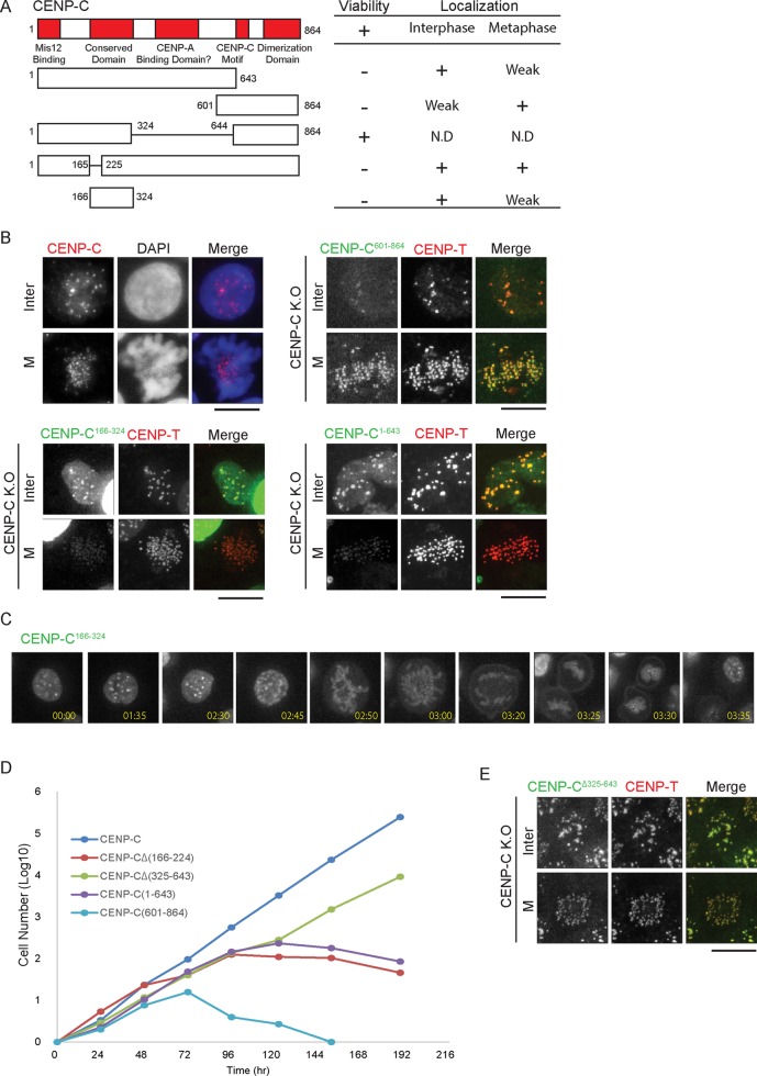
CENP-C166-324 and CENP-C601-864 localize to interphase and mitotic centromeres, respectively. (A) Schematic showing CENP-C domains (red) and the constructs used, along with their ability to rescue CENP-C depletion and localization throughout the cell cycle. (B) Representative images showing wild-type CENP-C (using an anti–ggCENP-C antibody), GFP-CENP-C601-864, GFP-CENP-C166-324, and GFP-CENP-C1-643 localization during interphase and metaphase. Kinetochores in CENP-C conditional-knockout cells expressing GFP constructs were visualized by CENP-T immunostaining at 36 h after addition of tetracycline. Bars, 10 μm. (C) Live-cell imaging of cells expressing GFP-CENP-C166-324. (D) Growth curves for CENP-C–deficient cells expressing the indicated CENP-C constructs. Tetracycline was added at t = 0, and the number of live cells was counted. (E) Localization of CENP-C∆325–643 and CENP-T in CENP-C–deficient cells expressing CENP-C∆325–643. Bars, 10 μm.

