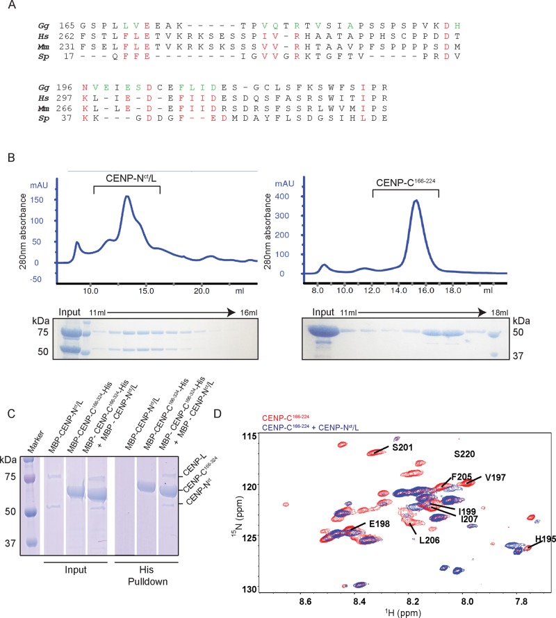
FIGURE 3:
Middle portion of CENP-C directly binds to the CENP-N-L complex in vitro. (A) Red letters show residues conserved between Gallus gallus (Gg), Homo sapiens (Hs), Mus musculus (Mm), and Schizosaccharomyces pombe (Sp). Green letters indicate residues possibly involved in the interaction between ggCENP-C166-324 and ggCENP-Nct/L (see D and Supplemental Figure S3). (B) Gel-filtration profiles for the CENP-Nct/L complex and CENP-C166-224. Peak fractions were visualized using SDS–PAGE. The CENP-Nct/L complex was prepared from MBP-CENP-Nct (54 kDa) and MBP-CENP-L (80 kDa). (C) Histidine pull down of CENP-C166-324 with the CENP-Nct/L complex. (D) 1H15N HSQC spectrum of free CENP-C166-224 (red) overlaid with that of CENP-C166-224 in complex with CENP-Nct/L (blue). Residues that showed shifted or abolished peaks are labeled.

