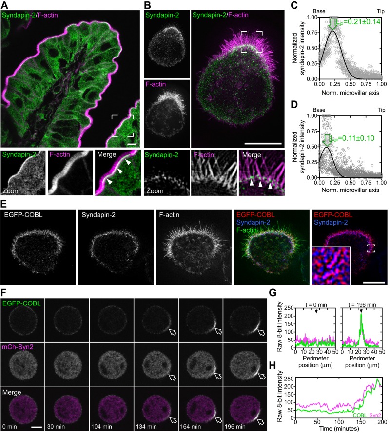
FIGURE 6:
Syndapin-2, similar to COBL, localizes to the base of BB microvilli. (A) Endogenous syndapin-2 localization (green) and F-actin signal (magenta) in a villus from human small intestine. Boxed corners indicate zoom region; arrowheads highlight syndapin-2 localization near the base of the BB. (B) SIM projection of endogenous syndapin-2 (green) in an Ls174T-W4 cell reveals enrichment at the base of the BB. Boxed corners indicate zoom region. Arrowheads highlight syndapin-2 localization at the base of the BB. (C, D) Line scans parallel to the microvillar axis (n = 25) show the distribution of endogenous syndapin-2 in human small intestine and Ls174T-W4 cells, respectively. Arrows highlight peak localization. (E) SIM projection of EGFP-COBL (red), endogenous syndapin-2 (blue), and F-actin (green) shows colocalization of EGFP-COBL and syndapin-2 at the base of the BB. Boxed corners in the red/blue channel overlay indicate the zoom region. (F) Time series montage of an Ls174T-W4 cell coexpressing EGFP-COBL and mCherry–syndapin-2 undergoing cell polarization and BB assembly. (G) Perimeter line-scan analysis of the cell shown in C at t = 0 and 196 min; black arrow in right-hand plot marks the position of the BB. (H) Intensity profile over time of a 2.5-μm2 region of interest at the base of the forming BB in F. Scale bars, 5 μm.

