
Figure 1.
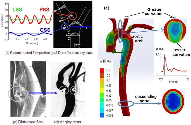
(a) Shear stress profiles at the lateral and medial walls of arterial bifurcations. (b) Pulsatile shear stress (PSS) occurs at the medial wall (red circle), whereas oscillating flow (OSS) occurs at the migrating stagnation point of the lateral wall (blue circle). (c) Flow separation and disturbed flow develops at the lateral wall. (d) Angiogram supports the predilection sites for atherosclerosis. (e) Spatial variations in wall shear stress profiles at an instantaneous moment in systole. The magnitude of wall shear stress is relatively high in the ascending aorta, greater curvature, and descending aorta. Cross-section at the aortic arch reveals an eccentric distribution of high shear stress in the greater curvature but low in the lesser curvature. Cross-section from the descending aorta reveals concentric high shear stress.