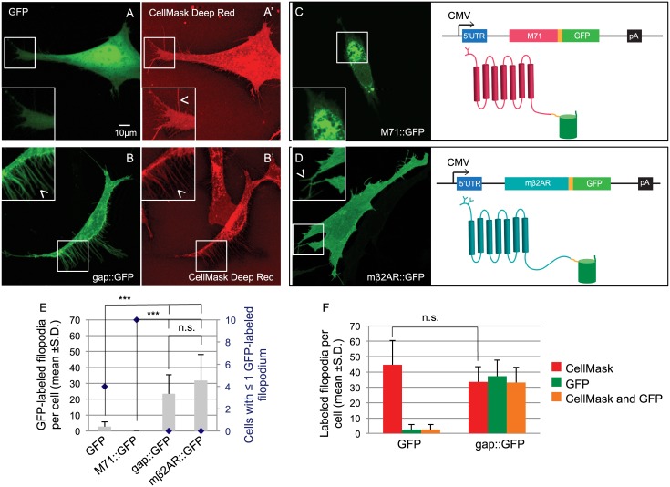
Fig 2. Expression of GFP and GPCR::GFP in OP6 cells.
(A) Cytosolic GFP shows diffuse fluorescence in the cytoplasm of an OP6 cell and does not co-localize with the plasma membrane staining CellMask Deep Red in filopodia (A’, arrow head in magnified image). (B) On the contrary, gap::GFP, shows a sharp edge staining of a whole OP6 cell and co-localize with the plasma membrane staining CellMask Deep Red in filopodia (B’, arrow head in magnified image). (C) The M71 coding sequence missing a stop codon (pink box) was cloned followed by a linker sequence (yellow box) and the coding sequence of GFP (green box). The peGFP-N1 backbone includes the cytomegalovirus promoter (CMV, arrow), a 5’ untranslated region (5’UTR, blue box) and a polyadenylation sequence (pA, black box). The resulting 2D structure of the fusion protein is showed, the M71 odorant receptor has a putative 7 transmembrane domains structure and is glycosylated at its N-terminus (pink diagram). The GFP protein, composed of eleven β-barrels, is fused to the Ct of M71 (green diagram) after a 9 amino acids linker (in yellow). M71::GFP shows intense perinuclear localization in an OP6 cell (see picture) and does not localize to filopodia. (D) Using the same strategy the mβ2AR sequence (teal box) was cloned in the same backbone. mβ2AR has also 7 transmembrane domains but 2 N-linked glycosylation sites located in its Nt (see 2D topology). When express in OP6 cells (see picture) mβ2AR::GFP stains homogeneously the whole cell and locates in filopodia (arrow head in magnified image), like gap::GFP. (E) Cells expressing GFP and M71::GFP have very low number of GFP-labeled filopodia (gray bars, 2.8 ±2.9 and 0.0 ±0.0 GFP-labeled filopodia per cell respectively), whereas cells expressing gap::GFP and mβ2AR::GFP show the same numerous number of filopodia. In addition many cells expressing GFP and M71::GFP show ≤1 GFP-labeled filopodium whereas all cells expressing gap::GFP or mβ2AR::GFP have more filopodia (blue diamonds, 2nd y axis in graph). (F) Counts of GFP- and CellMask Deep Red-labeled filopodia for cells expressing GFP or gap::GFP. The number of filopodia revealed by plasma membrane staining is the same for both type of cells (44.7 ±15.9 and 33.6 ±9.9 respectively), only in cells expressing gap::GFP-filopodia are also GFP-labeled (orange bars in graph, 2.6 ±3.2 and 33.2 ±9.8 co-labeled filopodia per cell respectively). All filopodia counts are represented as average for 10 cells ± standard deviation. ***means significantly different from mβ2AR::GFP, one-way ANOVA followed by Scheffe tests, p<0.001. n.s. means not significantly different from mβ2AR::GFP, p>0.001.

