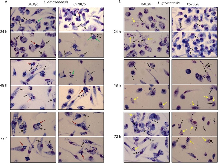
Fig 2. In vitro macrophage infection with L. amazonensis or L. guyanensis.
Peritoneal macrophages of BALB/c (left panel) or C57BL/6 mice (right panel) were infected with L. amazonensis or L. guyanensis. After the indicated time points cells were stained with May-Grünwald, followed by Giemsa, method. Images were obtained using QCapture Pro 7 Imaging Software (QImaging) obtained from http://www.qimaging.com/support/softwarereleases/030107_qcappro.php. Black arrows—amastigotes; green arrows—structures reminiscent of apoptotic bodies or condensed nuclei; yellow arrows—structures reminiscent of disintegrated cells; red arrows—structures reminiscent of apoptotic cells that seem to have been phagocytized by (or are attached in) other macrophages.

