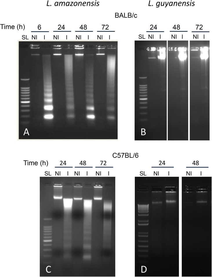
Fig 6. Macrophage DNA fragmentation pattern after in vitro infection with L. amazonensis or L. guyanensis as shown by agarose gel electrophoresis.
Peritoneal macrophages of BALB/c (A and B) or C57BL/6 mice (C and D) were infected or not with L. amazonensis (A and C) or L. guyanensis (B and D). DNA was extracted after the indicated time points and submitted to electrophoresis on agarose gel at 1.8%. SL—Step Ladder of 100 bp; NI—non-infected; I—infected.

