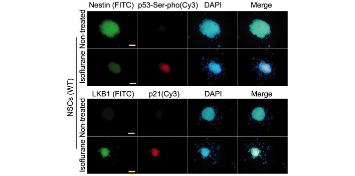
Fig. 3.
Immunofluorescence analysis of the Lkb1-p21 signalling pathway in WT NSCs. The protein expression levels of LKB1, p53-Ser-pho and p21 in isoflurane-treated WT NSCs were significantly increased compared with the untreated cells (scale bar, 50 µm). FITC, fluorescein isothiocyanate; DAPI, 4′,6-diamidino-2-phenylindole; WT NSCs, wild-type neural stem cells; p53-Ser-pho, Ser15-phosphorylated p53.

