
Figure 2.
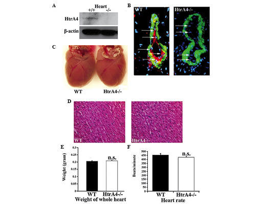
Expression of HtrA4 in the mouse heart. (A) Immunoblots were performed to determine the protein expression levels of HtrA4 and β-actin in heart extracts from WT and HtrA4−/− mice. (B) Immunofluorescence staining of HtrA4 and α-smooth muscle actin on cardiac tissue sections from WT and HtrA4−/− mice was performed. Arrows indicate endothelial cells (magnification, ×60). (C) Images of the heart from a 3-month-old HtrA4−/− mouse and its WT littermate were captured. (D) Hematoxylin and eosin staining of a cardiac tissue section from WT and HtrA4−/− mice (magnification, ×10) was performed. (E) The weight of the hearts from 3-month-old WT and HtrA4−/− mice were plotted in a histogram (n=6; P=0.40). (F) The heart rates in beats/min of the 3-month-old WT and HtrA4−/− mice were compared (n=15; P=0.17). n.s., non-significant; WT, wild-type; HtrA4, high temperature requirement factor A4.