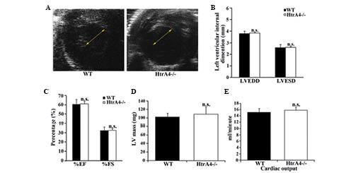
Figure 3.

Echocardiographic analysis of the cardiac function of HtrA4−/− mice. (A) Short-axis echocardiography demonstrated that the diameters of the left ventricles of the WT and HtrA4−/− mice were similar. The arrows show the size of the intraventricular diameters during diastole. (B) The LVEDD and LVESD of the left ventricles of the WT and HtrA4−/− mice were measured (n=6; P=0.73 and P=0.55, for LVEDD and LVESD, respectively). (C) The EF and FS of the left ventricles of WT and HtrA4−/− mice were determined (n=6; P=0.39 and P=0.34, for %EF and %FS, respectively). (D) The masses of the left ventricle from the WT and HtrA4−/− mice were measured and corrected by echocardiography (n=10; P=0.33). (E) The cardiac output of the WT and HtrA4−/− mice was determined (n=9; P=0.61). The results of these experiments were n.s. comparing WT with HtrA4−/− mice. EF, ejection fraction; FS, fractional shortening; LVEDD, left ventricular end diastolic diameter; LVESD, left ventricular end systolic diameter; HtrA4, high temperature requirement factor A4; n.s., non-significant; WT, wild-type.
