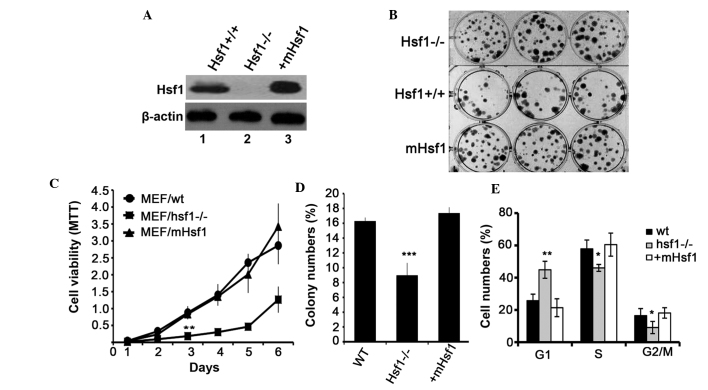
Figure 1.
Hsf1 knockout inhibits MEF cell proliferation. (A) Expression of Hsf1 proteins in the SV40/TAG-transformed MEF cell lines: Lane 1, MEF/wt; lane 2, MEF/Hsf1-/-; and lane 3, MEF/mHsf1. (B) Clone formation of the three MEF cell lines in flat cloning assay. (C) The growth viability of the three MEF cell lines in an MTT assay. (D) The quantification of colony-forming efficiency of the three MEF cell lines by flat cloning assay. (E) The effects of Hsf1 on the cell cycle of the three MEF cells. *P<0.05, **P<0.01, ***P<0.001, MEF/Hsf1-/- cells vs. MEF/wt and MEF/mHsf1 cells. Hsf1, heat shock factor 1; MEF, mouse embryonic fibroblast; SV40/TAG, simian virus 40/T antigen; wt, wild type; mHsf1, Hsf1 null MEF cells expressing mouse Hsf1.

