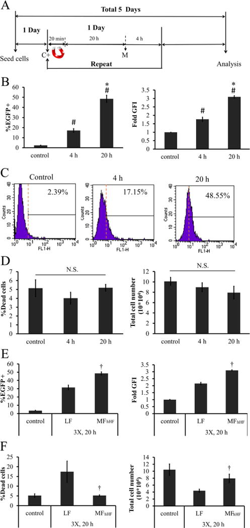
Figure 7.

Effects of MP:DNA incubation time on MF efficiency. (A) Timeline for multifection. C+: add MP:DNA complexes M: media change. (B—D) hHF-MSCs were incubated with MP:DNA for 4 or 20 h following withdrawal of the magnetic field: (B) transfection efficiency and GFI, (C) representative flow cytometry histograms, and (D) percentage dead cells and total cell count. (E,F) Comparison of optimized MF for hHF-MSCs (MFhHF) with the commercially available transfection reagent, Lipofectamine 2000 (LF): (E) percentage of EGFP+ cells and GFI, (F) percentage of dead cells and total cell number. The symbol # denotes p < 0.05 between nontransfected cells (control) and 4 or 20 h of incubation. The symbol * denotes p < 0.05 between 4 and 20 h incubation. The symbol † denotes p < 0.05 between LF and MFhHF. All values are the mean ± SD of triplicate samples in a representative experiment (n = 3). N.S.: not significant (p ≥ 0.05).
