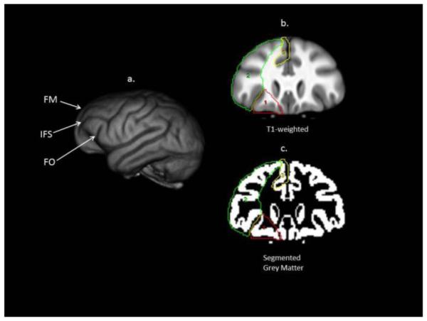
Figure 1.

Prefrontal cortex regions of interest. This figure illustrates: a) 3D rendering of a chimpanzee brain; b & c) coronal view of template brain and segmented grey matter volume with the three object maps corresponding to the prefrontal cortex, outlined in red (orbital), green (dorsal), and yellow (mesial). See text for specific description of the landmarks used to define the regions. FM = fronto-marginal sulcus; IFC = inferior frontal sulcus, FO = front-orbital sulcus. Note, white matter is included on the ROIs depicted on the T1-weighted scan but when the object maps were applied to the segmented grey matter volume, only voxels corresponding to gray matter were included in the calculation of volume.
