
Figure 1. Subbasal axons fully reinnervate the cornea and reform the vortex over time after trephine wounding but not after dulled blade and rotating burr wounding.
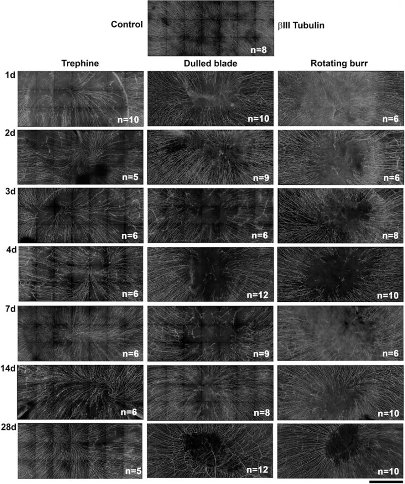
Representative stitched images of subbasal axons stained with βIII tubulin were taken by spinning disk confocal microscope to show the central 2 mm area of the mouse cornea encompassing the site demarcated by the 1.5 mm trephine in unwounded and corneas 1d, 2d, 3d, 4d, 7d, 14d, and 28d after wounding. The numbers of corneas used for quantification of reinnervation by Sholl analysis are indicated in the lower right corner of each image (Mag bar=500μm).