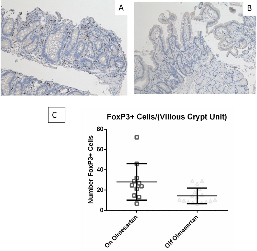
Figure 3.
FoxP3 expression in duodenum: Duodenal biopsies from a representative OAE patient while on (A) or off (B) olmesartan were stained with anti FoxP3 (brown). Panel C shows the mean with standard deviation of unpaired on and off samples. There was a statistically significant increase in the number of FoxP3+ cells with the use of olmesartan medoxomil (p<0.05), using the unpaired t test with Welch’s correction.

