
Figure 1.
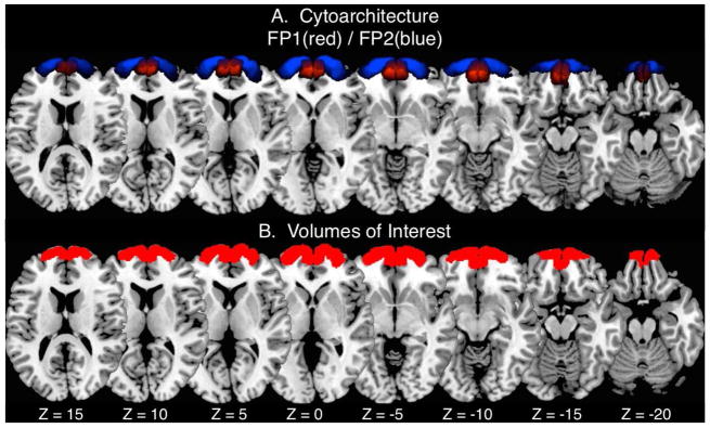
The volumes of interest used in the current CBP analysis were derived from A) the cytoarchitectonic borders of area frontopolaris one (FP 1 in red) and area frontopolaris two (FP 2 in blue) as identified in Bludau et al., (2014). B) The FP1 and FP2 within each hemisphere were joined to create a single binary mask for the right hemisphere and an additional binary mask for the left hemisphere for the CBP analysis.