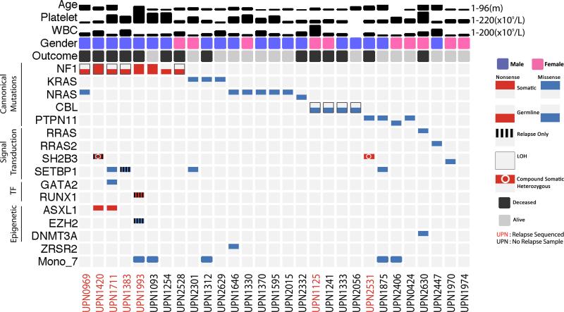
Figure 1.
Mutations identified by exome sequencing. Twenty-nine patients who underwent whole exome sequencing are displayed. Each patient is presented in a single condensed column including mutations identified at germline, diagnostic (noted in black) and relapse (noted in red) timepoints. Germline mutations are presented in colors in the bottom half of the box of any given gene and somatic mutations in the top half. Mutations only present at relapse are denoted with vertical striped bars. Loss of heterozygosity in a single gene is annotated with a thin black rectangle surrounding the mutation. Somatic compound heterozygous mutations are noted with a white circle.

