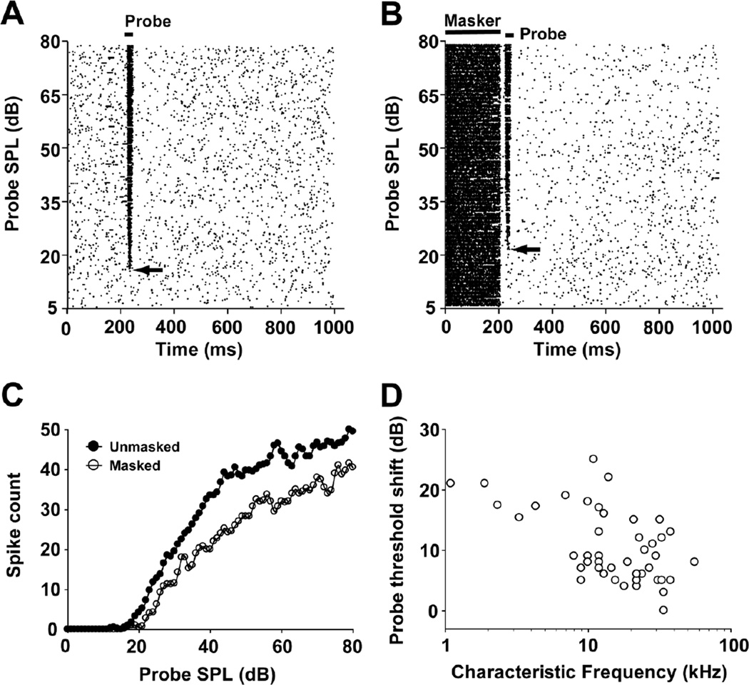
Fig. 3.
Example of unmasked (a) and masked (b) responses of a typical MNTB unit (neuron 13-02-1; CF=34 kHz; default masker condition: 200-ms CF tone presented at 57 dB SPL with a 10 ms masker-to-probe delay). Spike-time raster plots with probe intensity along the ordinate and time along on the abscissa. In this unit, the total number of spikes within the probe analysis window (see Fig. 1b) was determined from the probe threshold level to the maximum level tested under unmasked (probe threshold=17 dB SPL, total spikes = 2174) and masked (probe threshold=22 dB SPL, total spikes = 1588) conditions, respectively. Compared to the unmasked condition, the default masker caused a 27% decrease in spike count and a 5 dB increase in probe threshold (arrows). c Probe rate-level functions under unmasked (filled circles) and masked (open circles) conditions. d Threshold shift plotted over CF across the sample of 45 neurons, recorded under the default masker condition.

