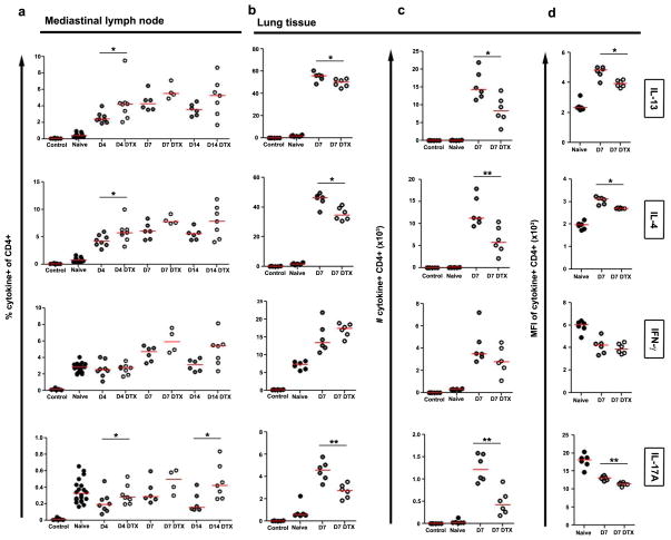
Figure 5. Reduced local but not systemic CD4+ Th2 cell responses underlie decreased lung inflammation and fibrosis.
(a) Mediastinal lymph node and (b) lung leukocytes isolated from naïve (lymph node n=20; lung D7 n=6), egg primed and challenged (lymph node D4 n=8; D7 n=6; D10 n=6; lung D7 n=6), and primed, challenged, and DTX treated (lymph node D4 n=8; D7 n=4; D10 n=7; lung D7 n=6) CD11b-DTR mice were restimulated with PMA plus Ionomycin and stained to compare cytokine producing capabilities of CD4+ T lymphocytes. (c) Total numbers of cytokine-producing inflammatory CD4+ T lymphocytes in the lungs, and (d) magnitude of cytokine production per cell was measured by the mean fluorescence intensity (MFI) of cytokine staining. Little differences in effector CD4+ T lymphocytes were observed in lung-draining lymph nodes. In contrast DTX treatment reduced the frequency, number, and intensity of IL-13, IL-4, and IL-17A-producing CD4+ effector T lymphocytes in the lungs. Data are presented as medians. Statistical significance was calculated using Mann-Whitney U test. * p<0.05, ** p<0.01. Results represent two independent experiments.

