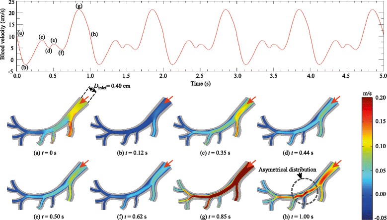
Fig. 3.

(Top) The physiological waveform used in this study was based on the work of Matsuo and co-workers [38], measured by means of a bidirectional Doppler flow meter catheter. (Bottom) The inner diameter of the inlet (right) is D inlet=0.40 cm. The velocity magnitude (represented by colour bar) in the simulated arterial flow corresponds to the velocity profile illustrated in the top figure. The red arrows denote the direction of flow at the inlet
