Fig. 5A–F.
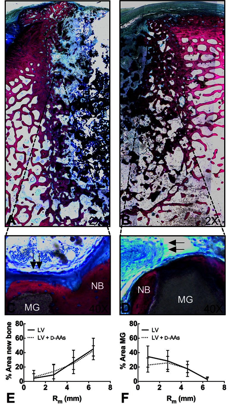
Half-view (of entire slide analyzed) low- (×2) magnification images of histologic cross-sections of defects filled with (A) low-viscosity (LV) or (B) LV+ d-amino acids (d-AAs) at 16 weeks show active remodeling. Sections were stained with Stevenel’s Blue and Van Gieson. Corresponding high- (×40) magnification images of highlighted portions of defects filled with (C) LV or (D) LV+ d-AAs show residual MASTERGRAFT® (MG) ceramic particles, new bone (NB), and vascular development (arrows). Area % (E) new bone and (F) MG at four regions in the defect measured by histomorphometric analysis show minimal differences in new bone formation or MASTERGRAFT® degradation between LV and LV+ d-AAs. Values are mean ± SD of eight samples per region. Statistical significance was determined by two-way ANOVA and Tukey’s multiple comparison test, post hoc.

