
Figure 4.
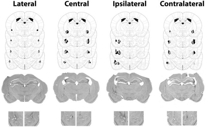
Minimum (gray) and maximum (black) accepted lesions for subjects with bilateral LA or CeA lesions as well as ipsilateral or contralateral LA-CeA lesions. Figures were adapted from Paxinos and Watson (2013) and profile damage 1.72, 2.16, 2.64 and 3.12 mm posterior to bregma. Representative images for each surgical condition are presented below the schematics with an enlargement of the amygdala region from each image presented below.