Figure 1.
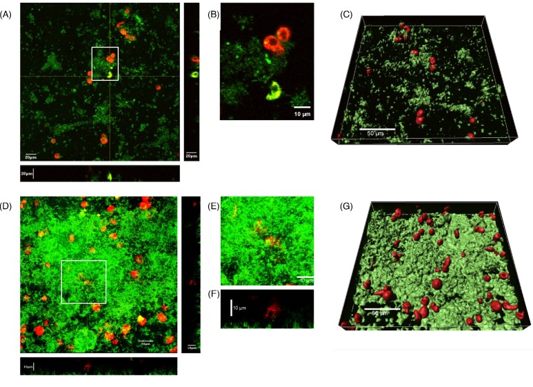
dHL60 cells do not enter established B. multivorans LMG 13010 and B. dolosa LMG 18941 biofilms. Bcc bacteria (1 × 10⁶ cfu ml−1), harboring a GFP-expression plasmid, were inoculated into flowcells and cultured under continuous throughput of defined, minimal medium at 37°C for 72 h. To the flowcell was added 4 × 10⁵ dHL60 cells ml−1, which had been stained with 5 μM Celltracker Red cytoplasmic dye (Molecular Probes), 30 min prior to image collection. Composite confocal micrographs of sections through the z-axial plane of biofilms formed by (A) B. dolosa LMG 18941 and (D) B. multivorans LMG 13010 were then generated, with biofilms proximal to the viewer. (B) Enlargement of a single micrograph of the area demarcated by white box in A showing a phagocytosing cell. (E) Enlargement of the area demarcated by white box in D to highlight (F) a transverse section corresponding to image E illustrating a cell resting above the biofilm. Using the data presented in A and D, discrete surfaces were overlaid onto the detected fluorescence intensities for each channel using Imaris software (Bitplane) in order to illustrate the spatial relationship between (C) B. dolosa and (G) B. multivorans biofilms, and dHL60 cells.

