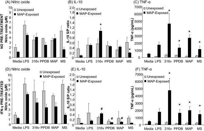
Figure 3.
Nitric oxide and cytokine responses of monocyte-derived macrophages to mycobacteria and antigens. PBMCs were isolated from the whole blood of unexposed control (n = 5), MAP-exposed (n = 10) cattle and monocytes isolated by CD14+ selection using MACS®. Cultured bovine macrophages were incubated for a period of 48 h with LPS (25 ng/mL), MAP 316v antigen (316v) or M. bovis antigen (PPDB; 100 μg/mL), live MAP or M. smegmatis strains at an MOI of 1:1 (viable count). Cells were cultured without pre-treatment (top panels A–C) or after IFN-γ pre-treatment for 1 h (lower panels D–F). Supernatants were assayed for production of nitric oxide (A, D); IL-10 (B, E) or TNF-α (C, F). Data are the mean plus standard error of the mean of three replicate cultures from one out of two independent experiments. Asterisk denotes a significant difference (P ≤ 0.05) in macrophage responses between the unexposed and MAP-exposed cattle. Hash denotes a significant difference (P ≤ 0.05) between the macrophage responses from MAP-exposed cattle without IFN-γ pre-treatment and after IFN-γ pre-treatment.

