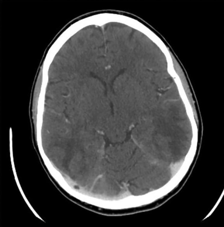
Figure 1.

CT head with contrast showing diffuse areas of abnormal low attenuation mostly involving the subcortical white matter of the medial frontal and parietal lobes as well as the subcortical and deep white matter of the occipital and posterior temporal lobes consistent with PRES.

 This work is licensed under a