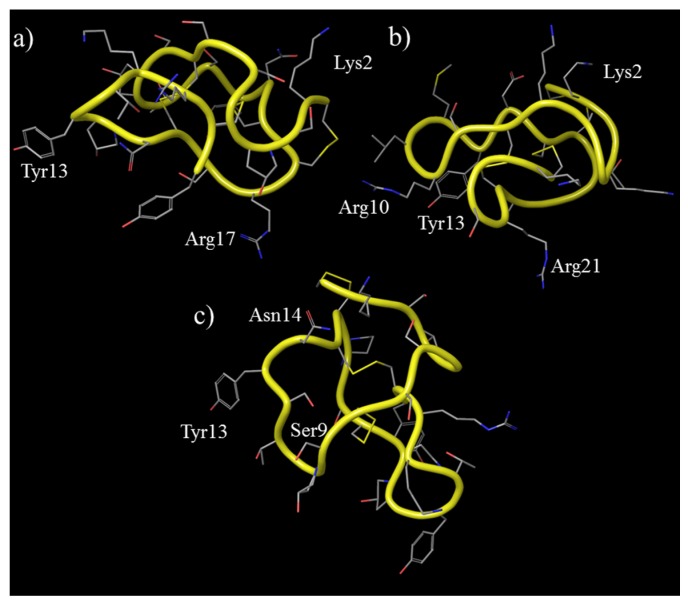
Figure 2.
Three-dimensional structures of ω-conotoxins that are relevant to this review. The peptide backbone structure is shown as a yellow tube, the side chain residues are shown as thin tubes and are colored according to the atom type, and hydrogen atoms are not shown. The amino acid side chains thought to be important for biological activity are highlighted. (a) ω-conotoxin GVIA (PDB code: 2cco); (b) ω-conotoxin MVIIA (PDB code: 1ttk); and (c) ω-conotoxin CVID (coordinates obtained from reference [29]).

