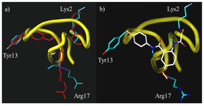
Figure 7.
Backbone structure of ω-conotoxin GVIA (yellow) with the amino acid side chains Lys2, Tyr13, Arg17 shown in teal; (a) overlaid with a model of the benzothiazole mimetic 8 [28]—Reproduced with permission; and (b) overlaid with X-ray crystal structure of the anthranilamide scaffold core without side chain mimics. The α,β-bond vectors are shown in purple and the bonds mimicking the α,β-bond vectors are shown in yellow [47]—Reproduced with permission.

