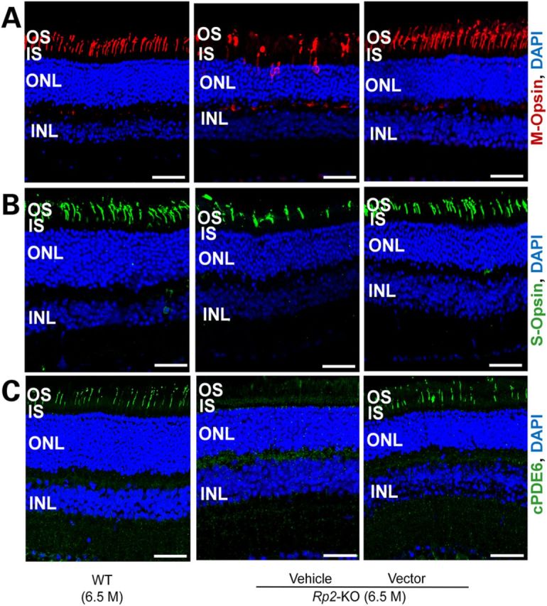
Figure 6.

Correction of cone opsin mis-localization and restoration of cone PDE6 expression in Rp2-KO retina following vector treatment. Immunostaining of M-opsin (A), S-opsin (B) and cone PDE6 (C) of retinal sections from a 6.5-month-old Rp2-KO mouse that received vehicle or 5 × 10⁷ vg AAV8-scRK-hRP2 vector administration at 4 weeks of age. Retinal sections from an age-matched WT C57/Bl6 mouse were used as controls. While M-opsin is mis-trafficked to OS, IS, nuclei and synaptic terminals in vehicle-treated retina, it is only localized to OS in vector-treated retina, similar to the WT mouse. Diminished cone PDE6 was observed in the vehicle-treated retina, whereas in the vector-treated retina, cone PDE6 restored and is localized to OS. Scale bar 50 μm. OS, outer segments; IS, inner segments; ONL, outer nuclear layer; INL, inner nuclear layer.
