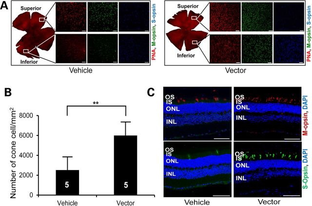
Figure 7.
Long-term preservation of cone photoreceptors in Rp2-KO mice following vector treatment. (A) Immunofluorescence analysis of retinal whole mounts of an 18-month-old Rp2-KO mouse that received subretinal administration of vehicle or 1 × 10⁸ vg AAV8-scRK-hRP2 vector at 4 weeks of age. Quantification of cone cells in retinal whole mounts (B) revealed a significantly higher number of cone cells in vector-treated eyes compared with vehicle-treated control eyes. The number of mice tested in each group is indicated by white lettering inside each bar. Data from vector- and vehicle-injected eyes were compared by two-tailed paired t-test and represented as mean ± SEM. **P < 0.01. (C) Retinal sections showing a higher number of S- and M-opsin expressing cells in vector-treated eye than in vehicle-treated eye. Scale bar 50 μm. OS, outer segments; IS, inner segments; ONL, outer nuclear layer; INL, inner nuclear layer.

