Figure 8.
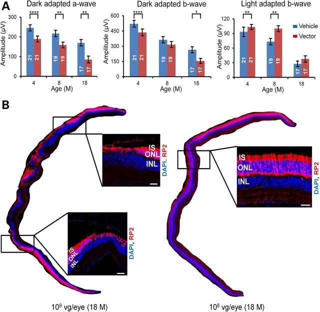
Retinal toxicity at high vector dose. (A) Dark- and light-adapted ERG responses at different time points in Rp2-KO mice treated with AAV8-scRK-hRP2 vector. Mice were injected unilaterally with 1 × 10⁹ vg/eye of the vector when they were 4–6 weeks old, with the vehicle injected to the contralateral eyes as controls. The number of mice tested in each group is indicated by white lettering inside each bar. The stimulus intensities for dark- and light-adapted ERGs were −4.0 to 3.0 and −1.0 to 2.0 log cd s/m2, respectively. Only the amplitudes with the highest flash stimuli are shown. The ERG response with a full range of flash stimuli is shown in the Supplementary Material, Figure S11. ERG amplitudes from vector- and vehicle- injected eyes were compared by two-tailed paired t-test and represented as mean ± SEM. *P < 0.05, **P < 0.01, ****P < 0.0001. Lower dark-adapted ERG response in the vector-treated eyes indicates retinal toxicity likely caused by high RP2 expression. (B) RP2 staining of retinal sections from 18-month-old Rp2-KO mice that received injections of 1 × 10⁹ or 1 × 10⁸ vg/eye of the vector. Thinning of ONL was observed at multiple regions in the retina treated with 1 × 10⁹ vg/eye, while relatively normal ONL thickness was maintained in the 1 × 10⁸ vg-treated retina. The magnified images of the marked areas are shown. Scale bar 50 μm. IS, inner segments; ONL, outer nuclear layer; INL, inner nuclear layer.

